Sunday, October 24, 2021

Slow and scary, deadly scatteredly.

 If there’s a word that is synonymous with cancer, it’s chemotherapy.  It’s what most people have in their mind when they are talking to a cancer patient…… ‘have you had chemotherapy‘ or ‘when do you start chemotherapy‘.


An acquaintance asked nonchalantly some time ago ‘how did you get on with chemotherapy’ – he was surprised to hear my daughter hadn’t had it despite her widespread disease.  Cue – lengthy explanation!  I wasn’t annoyed by the question; I just think people automatically assume every cancer patient must undergo some form of systemic chemotherapy.  If you read any newspaper article about cancer, they do nothing to dispel that myth, as many articles contain a story about a cancer patient with no hair.


Sure, chemotherapy is not the nicest treatment to receive, and it does have pretty awful side effects for most. I, watching my daughter go through 3 or 4 months of this treatment where she seemed to be confined to a combination of her bedroom and her bathroom.  And it did shock me to see her without hair after radiation therapy.  I would never want anyone to go through that and it really brings it home when it happens to a close member of your family.


Despite its terrible news 📰 in regard to toxicity and its awful side effects, chemotherapy is widely used in many cancers.  Statistics show that it does work for many patients even my daughter  was pushed to it . However, I suspect it has a limited future as more efficient and less toxic drugs and delivery systems come online downstream. 

Biopsy is the gold standard to confirm the diagnosis but does not differentiate between pheochromocytomas and paragangliomas. Since most paragangliomas are vascular, a preoperative biopsy is not common.

Reside in ignorance. Where ignorance is the Master They handover prescription for disaster; If you don't know either you loose your life being a scapegoat or be blocked by doctors who are supposed to keep themselves updated and save the lethal Neuroendocrine Cancer warrior fighting since 3 years old of a genetic mutation One is named as king of the sky, the doctor of hormones Went into warzones, the next whose negligence pushed me to last stage is always proud and egotistical about mastery, wealth, superiority, this she's most desired. As for her eminence.the protegee smelly as my poop akin to perfume


Immunotherapy is often used as a replacement for chemotherapy, but this may be a while yet.  So, for now, millions of cancer patients worldwide will continue to be prescribed chemotherapy as part of their treatment regime. What if the patient is immunocompromised?


However, for some cancers, chemotherapy is not particularly effective. Neuroendocrine Tumours (NETs) is one such cancer, particularly for the low-grade of well- differentiated tumours, although but can be more effective in particular anatomical locations and at the higher numbers in Grade 2 and in Grade 3 well differentiated NET. It’s a first line therapy for most poorly differentiated Neuroendocrine Carcinomas.   I’m not saying the lower grades will never receive chemotherapy – that door is always left open for those with progressive cancer who perhaps have run out of treatment options. 


Putting Grade 3 to one side, I’ve heard people say that NETs is the ‘good‘ cancer or the ‘good looking’ cancer often citing the chemotherapy thing as some justification. That is of course a stupid thing to say.  I accept that not everyone will lose their hair and not every chemo will cause hair loss.


Here’s the message.  Many other treatments come with pretty challenging side effects. Moreover, the side effects and the consequences of these other treatments can last for some time, and for many, a lifetime. For example, with NETs:


Surgery can be pretty extensive, in some cases radical and life changing.  Many cancer patients receive surgery for NETs which is still the only real ‘curative’ treatment, although for most, it’s cyto reductive or palliative in nature.  If you lose bits of your small intestine, large intestine, liver, spleen, cecum and appendix, gallbladder, stomach, rectum, lungs, pancreas, thyroid, parathyroids, pituitary gland, adrenal gland, thymus gland, ovaries, oesophagus (…….I could go on), this comes with various side effects which can present some quality -of - life issues.  There can be huge consequences of having this treatment.


Other ‘consequences’ of cancer surgery include (but are not limited to), pulmonary emboli (blood clots), lymphedema, short bowel syndrome, gastrointestinal malabsorption, diabetes.


Somatostatin Analogues do a great job, but they do add to some of the effects of surgery (e.g. malabsorption).


Even the so-called ‘silver bullet’ treatment Peptide Receptor Radio Nuclide Therapy (PRRT) can have pretty severe side effects and presents some risk to kidneys and bone marrow as a long-term consequence.

I am adviced this


The thyroid gland is not visualised (post thyroidectomy). Coalescing enhancing nodular areas are noted in the location of right lobe of thyroid, abutting the right lateral and anterior wall of trachea. The confluent lesion in this location measures 2.7 x 2.3 x 1.5cm in size. A smaller enhancing area is noted in the location of left lobe of thyroid, abutting the tracheal wall, measuring 1.0 x 0.7 x 0.6cm in size. Multiple calcified nodular lesions are seen in both lung fields, relatively larger and more in number in the upper lobes, suggestive of old granulomatous lesions. Fibrotic bands are also noted in both lungs. Air trapping is noted in both lung fields. Few small thin walled cysts are also noted in both lungs. Diffuse inhomogenous mosaic pattern enhancement is noted in the liver parenchyma in arterial phase Right kidney is normal in position and size. Few small subcentimeter sized cysts are noted in right kidney, the largest in its upper pole measuring 0.9 x 0.8cm. The largest cyst in upper pole of right kidney shows relatively higher attenuation in non contrast sections, suggestive of a complex cyst (Bosniak type 2). There is no hydronephrosis or ureteric dilatation. Left kidney is normal in position and size. Areas of cortical scarring are noted in left kidney, more prominent in the upper polar region. A simple cyst is seen in its mid polar region in peripelvic location, measuring 2.7cm in diameter. Another small subcentimeter sized simple cyst is also noted peripherally in its mid polar region. There is no hydronephrosis or ureteric dilatation. A hypodense mass is noted in uterus, which may represent a fibroid. The thyroid gland is not visualised (post thyroidectomy). Coalescing enhancing nodular areas are noted in the location of right lobe of thyroid, abutting the right lateral and anterior wall of trachea. The confluent lesion in this location measures 2.7 x 2.3 x 1.5cm in size. A smaller enhancing area is noted in the location of left lobe of thyroid, abutting the tracheal wall, measuring 1.0 x 0.7 x 0.6cm in size. CT study reveals focal enhancing areas in the location of right and left lobes of thyroid gland, relatively larger on right side. These are seen in the last PET-CT study of 07/02/2022 Multiple calcified nodular lesions are seen in both lung fields, relatively larger and more in number in the upper lobes, suggestive of old granulomatous lesions. Fibrotic bands are also noted in both lungs. Air trapping is noted in both lung fields. Few small thin walled cysts are also noted in both lungs. No significant enlargement of mediastinal/hilar lymph nodes is seen. Few small subcentimeter sized calcified mediastinal lymph nodes are noted, consistent with old granulomatous lesions. Trachea and main stem bronchi appear normal. OPINION: CT study reveals multiple calcified nodular lesions in both lungs, more prominent in the upper lobes, suggestive of old granulomatous lesions. Few small calcified mediastinal lymph nodes are also noted. Compared to last PET-CT of 07/02/2022, there is no significant interval change. Please correlate clinically. Spleen is normal in size. Multiple small calcific nodular foci are noted in spleen, suggestive of old granulomatous lesions. Both adrenals appear normal. Right kidney is normal in position and size. Few small subcentimeter sized cysts are noted in right kidney, the largest in its upper pole measuring 0.9 x 0.8cm. The largest cyst in upper pole of right kidney shows relatively higher attenuation in non contrast sections, suggestive of a complex cyst (Bosniak type 2). There is no hydronephrosis or ureteric dilatation. Left kidney is normal in position and size. Areas of cortical scarring are noted in left kidney, more prominent in the upper polar region. A simple cyst is seen in its mid polar region in peripelvic location, measuring 2.7cm in diameter. Another small subcentimeter sized simple cyst is also noted peripherally in its mid polar region. There is no hydronephrosis or ureteric dilatation. Diffuse inhomogenous mosaic pattern enhancement is noted in the liver parenchyma in arterial phase. CT study reveals multiple homogenously enhancing intracranial extra-axial dura based nodular lesions. Compared to last PET-CT of 07/02/2022, there is no significant interval change. Please correlate clinically. -- Test Name:CECT HEAD,CECT NECK,CT CHEST WITH CONTRAST (CECT CHEST),TRIPLE PHASE CECT WHOLE ABDOMEN CECT NECK,CT CHEST WITH CONTRAST (CECT CHEST),TRIPLE PHASE CECT WHOLE ABDOMEN TRIPLE PHASE CECT WHOLE ABDOMEN Anybody willing to help?


Daughter had  not had chemotherapy and I would rather avoided it if I can. However, as I’ve hinted above, there are other harsh (….perhaps harsher?) treatments out there. Moreover, whilst hair normally grows back, your small intestines, lungs and pancreas won’t.  Many people will have to live for the rest of their life with the consequences of their cancer and its treatment.


It sometimes appears that every other cancer article involves someone undergoing chemotherapy.  I just wish someone would write an article about my daughter's small intestines and ascending colon, the malabsorption issues as a consequence of that, my missing mesenteric lymph nodes, my retroperitoneal fibrosis, not forgetting to mention my diseased liver, my left axillary lymph nodes (and the mild lymphedema I now have after their removal), her right supraclavicular lymph nodes, thyroid lesion and my daughter's hypoparathyroidism and hyperthyroidism, there may be due to that, my small lung nodule, rib lesion and pulmonary emboli or blockages which after nearly 6 years of daily blood thinning injections made the abdomen look and feel like she'd done rounds with Mary Kom who already had a couple of rounds making her body shake and she broke cups and plates and spilt water , juice and mouthwash. However, it just wouldn’t be a good picture, nor would it be as powerful as one of a person with no hair or just partial hair in the right side. Just saying!


I look well, I still have all my hair – but you should see my insides!

insides.


“Patients present in an almost surrealistic manner – a mix of tumor- related symptoms and signs, with bizarre and sometimes grotesque endocrine syndromes”


The article from 1987, when somatostatin analogues were starting to be used, must have been a hugely exciting time, perhaps more so than when peptide receptor radiotherapy (PRRT) became a thing in the 21st Century. 

In his own paper, Dr Lewis quite rightly points out the continued confusion whereby the classification of neuroendocrine tumors (NETs) has long been obfuscated by the etymology of the German word karzinoide, a misnomer if it is translated to mean that this cancer - like disease is uniformly benign. Clearly there’s still work to do to get rid of that obfuscation.


Covering the issue of patient quality of life.  Clearly there’s a balance required making therapy decisions often tough for the physician and patient, particularly when the therapy may be different across the broad spectrum of NET types and primary locations. Another area where many unmet needs still remain.


And he approaches the tricky subject of therapy sequencing with the words “This risk - benefit calculus is crucial in caring for patients whose longevity can be measured in years, if not decades, and for whom therapeutic options have multiplied to the point that the sequence of their use must be considered“. Working out the best sequencing is difficult, particularly with so many factors involved, not least the different types of NET and primary locations.  This is really a work in progress and perhaps will always be.


https://youtu.be/JrtgufwSISQ


Maryfilomena E Rita Viñcenza I am honoured by daughter's comment "Payel Bhattacharya you are what I call a beautiful soul . You see, I knew !!!! I see such light coming from you . There is a special reason why God saved you and uses you for his glory . I am so honored to have Met you . May God bless you . You have lightened up my mind and spirit ; you see , it is 20 years from 9/11, my love of my life Til death did we part , Mark , died from being a first responder a NYPD Sgt , who got kidney cancer from working months there . This puts my gift of life into perspective once again . I like you always chose to find the flowers the birds the breeze the music in life in the lil moments of the day . I have lost so much of my light since Mark died nearly 8 years ago . This is how I have written poems in the past and more importantly how I have spoken to encourage and enlighten others in their dark moments . Thank you . I’ll pray blessings over you . 💝"


I am happy she has  a great friend too.
Friendship is one of life's greatest treasures.
Souls connected in space and time beyond any measure 

Even if we don't see each other for years

Emotions yet untold;

As time goes on, and bonds grow strong,

They will all unfold.

You are my daughter's great friend  and I am happy.
 Pray she has such friends who care for her survival 
Not good to be bickering, having ego when it concerns a life.
  Note from Payel Bhattacharya's mother


That's what I learnt in life. It's about willing to hold your hands when you need them most. I let those creatures walk away who didn't want to stay because my destiny is not tied to them.
Apart from the couple of egotistical doctors my endocrinologist blocked me for maximum security while I have stage IV metastastatic neuroendocrine cancer. says my daughter

Next week starting treatment in a new and good hospital with good doctor

Those who have a willingness to burst less Chinese crackers, have gala parties, save someone's life kindly donate. Last night too couldn't sleep because I am a Bengali and had only chicken broth.


Friday, October 22, 2021

Guruma and shishya tactical medical strategies where treatment is according to normal business strategies

"Somatostatin receptors may be over expressed in a myriad of diseases. Does not necessarily mean you have metastatic NET. And whether PRRT is the treatment for you or not needs to be assessed."
Said a medical professional with the thought in mind

We are the capable men we have a lot of ego

Who are you? A mosquito? You can't be bigger than my ego


Little time left and so much to accomplish

It's a moment to establish

But no one understands the eternal truth. 


Nor seek, for this is also sooth,


To hunger fiercely after truth

No one would ever know what it had cost have to accomplish 

Von Hippel-Lindau disease, or VHL disease, is one of more than 7,000 known inherited rare disorders. It is a disease caused by a mutation in the VHL gene, which normally prevents tumor growth. The mutation to this gene prevents it from working properly, resulting in both benign tumors and malignant tumors. The latter can spread and become metastatic. In people with VHL, tumors may develop in up to ten different parts of the body. All of these tumors involve the abnormal growth of blood vessels. Most of these tumors are benign, meaning that they will not spread to nearby organs. However, VHL tumors in the kidneys, adrenals, and pancreas can grow to a stage where they become “malignant,” where the cancer can spread to other parts of the body. The VHL Active? Patients and doctors to actively work together to prevent most of the worst effects of VHL.


 Neuroendocrine tumors overexpress somatostatin receptors, which serve as important and unique therapeutic targets for well-differentiated advanced disease. This overexpression is a well-established finding in gastroenteropancreatic neuroendocrine tumors which has guided new medical therapies in the administration of somatostatin analogs, both “cold”, particularly octreotide and lanreotide, and “hot” analogs, chelated to radiolabeled isotopes.

 The binding of these analogs to somatostatin receptors effectively suppresses excess hormone secretion and tumor cell proliferation, leading to stabilization, and in some cases, tumor shrinkage.

 Radioisotope-labeled somatostatin analogs are utilized for both tumor localization and peptide radionuclide therapy, with 68Ga-DOTATATE and 177Lu-DOTATATE respectively. Benign and malignant pheochromocytomas and paragangliomas also overexpress somatostatin receptors, irrespective of embryological origin.


 The pattern of somatostatin receptor overexpression is more prominent in succinate dehydrogenase subunit B gene mutation, which is more aggressive than other subgroups of this disease. While the Food and Drug Administration has approved the use of 68Ga-DOTATATE as a radiopharmaceutical for somatostatin receptor imaging, the use of its radiotherapeutic counterpart still needs approval beyond gastroenteropancreatic neuroendocrine tumors. Thus, patients with pheochromocytoma and paraganglioma, especially those with inoperable or metastatic diseases, depend on the clinical trials of somatostatin analogs. The review summarizes the advances in the utilization of somatostatin receptor for diagnostic and therapeutic approaches in the neuroendocrine tumor subset of pheochromocytoma and paraganglioma; we hope to provide a positive perspective in using these receptors as targets for treatment in this rare condition.

 In a known case of VHL syndrome; PET/CT scan findings reveal multiple DOTANOC avid( somatostatin receptor expressing) dural based nodular and plaque-like lesions as described.

It all started with multiple hypodense areas near neck and body region of pancreas.


Inference
Hypoechoic nodules show significant vascularity in the jugular region shows a Doppler 
FDG avidity in lungs --- VHL is associated with benign cysts in the lungs. When first noted at the National Institutes of Health, biopsies were performed. All of the lesions were benign, with no metastases from other organs. The presence of a lung hemangioblastoma is a possible rare manifestation in VHL patients.
My lungs have been stated as the previously fibrotic and fibrocalcific changes... in bilateral lung apices fibronodular lesions showing specks of internal calcification in bilateral lungs has been copied in several reports. Never looked at the details.


Lung carcinoids are rare tumors, although the incidence has increased during the  last decades. Lung carcinoids may be located centrally (60–84 %) in the main or lobar bronchi or  peripherally. Central carcinoids may be seen at bronchoscopy as a polypoid, highly  vascular, cherry-red, white-yellow, or brownish-yellow endobronchial tumor . The tumor often infiltrates the surrounding lung parenchyma. Peripheral  carcinoids, which are located in segmental bronchi or more distally, are not accessible by bronchoscopy. They may be multiple and surrounded by small satellite. lesions, tumorlets. Typical carcinoids may be located anywhere in the lungs, while  atypical carcinoids are more often peripheral.


Neuroendocrine tumors (NETs) are a group of heterogeneous cancers arising from a variety of anatomic sites. Their incidence has increased in recent years. This study aimed to analyze the prognosis of NETs originating from different anatomic sites.

NETs usually express somatostatin receptors; therefore, somatostatin expression  can be used both diagnostically and therapeutical. Somatostatin receptor imaging ( 111 In-DTPA-octreotide or preferably  68 Ga-DOTATATE) can be used for initial  staging, follow-up and selecting patients for peptide receptor radionuclide therapy  (PRRT). According to the localization of the primary tumour, computerised  tomography and/or magnetic resonance imaging (MRI) should also be used for diagnosis and staging. Ultrasound and endoscopic ultrasound can be used when necessary  as complementary examinations.  (FDG)-positron emission  tomography (PET) can be used in diagnosis, staging and follow- up of aggressive,  poorly differentiated NETs. FDG-PET may also show transformation to aggressive  biological behaviour. The role of newer PET tracers warrants further validation for routine clinical practice. Endoscopic evaluation of the patients with esophagogastroduodenoscopy, colonoscopy, double- balloon enteroscopy or capsule endoscopy  is critical according to the localization of the primary tumour and also in patients with  primary unknown metastatic  neuroendocrine tumour or carcinoma

 "Somatostatin receptors may be over expressed in a myriad of diseases. Does not necessarily mean you have metastatic NET. And whether PRRT is the treatment for you or not needs to be assessed" as told by a doctor was a reputable breeder mulesing, De clawing,der knocking with an ugly practice of traveling injuring and killing seemed bad hence harvesting in one Bengali word " puschi" she indicated we are not humans but animals, abusive to my mother She was under the illusion we are staying for 20 years  in Delhi and has done several Dotanoc whereas only once before RCC it was done to inspect if it's adrenal pheochromocytoma also about and supporting us for years. 2015 was the RCC year but FDG pet Scans we're supposed to be done every 6 months with contrast given in  every alternate scans but I never received any contrast ear blasting retort was you had to pay for it yet payment for contrast was never asked for in the guise of a do-gooder calling my mum "Mashima". Telling me your mother deserves more respect". It's difficult to discern the person behind a mask. 


Computed Tomography Scan (CT) scans were used in the past for abdominal imaging. The problem is radiation exposure. Use of contrast agents has resulted in reduced radiation, while maintaining image quality. She had used contrast once after RCC that's all

Doctors may describe a NET by its World Health Organization (WHO) grade (G). Knowing the grade helps the doctor create a treatment plan for the NET. "Grade" and "stage" may be mentioned at the same time while NETs are being discussed, but they are very different elements in understanding the risk a tumor poses to a person. This section covers grading for NETs. The WHO grades for NETs include:

  • Grade 1 (low-grade tumor): These cells divide at a low rate and therefore grow slowly.

  • Grade 2 (intermediate-grade tumor): These cells divide at an intermediate rate.

  • Grade 3 (high-grade tumor): These cells divide at a fast rate and therefore grow quickly.


The doctor's protégé took the warfare
ahead when action hospital was approached who did her duty

"NET grade please" " Report with grade of NET" " A pathological diagnosis of NET with grade of tumor is needed first" " A biopsy is done in hospitals or lab setups for a pathological diagnosis. Your treating team needs to advice for it and plan the site of biopsy."


Because I told her what her mentor told me:


 "Payal...either you can play doctor or I can..."
 "If you want my advice or support..you cannot dictate what test or scan I do and how"
"You want to get any test you want..pay for it and get it done wherever you want"
Let me know when you have the pathology report for "NET diagnosis with grade of tumor.
 I am available in hospital everyday. You can get in touch when you have the reports for an appointment.
 I am at work. Please message me when you need to take an appointment. 
For any emergency or other advice, it's best if you contact your treating team."
They had perhaps passed med school by copy pasting
Not understanding just memorizing.

"pheochromocytomas/paragangliomas are highly vascular and may be either parasympathetic or sympathetic. I don't find sense doing biopsy of a highly vascular tumour" and blocking me when I denied to die by their experiment tomorrow.... where there is vascularity seeding is supposed to happen and more cancer hence not even a couple of years like Irrfan Khan


Through life's hard task

 I did not ask

 Try finding out the man behind the mask

Because masks that hide, masks that reveal is a disagreeable, dreadful task


The whole world acting out a farce

All  turning into a farce

Promis'd which dwindled to a farce?

In the darkness sunlight became sparse

Spitting out filthy words

Under the influence of a silk purse


  • Gallium 68 (68Ga) (DOTA)–octreotate (DOTATATE, GaTate) positron  emission tomography (PET)/computed tomography (CT) is an  imaging technique for detecting and characterizing neuroendocrine tumors (NETs). GaTate, a somatostatin analog, has recently  been accorded orphan drug status by the U.S. Food and Drug  Administration, thereby increasing interest in and availability of this  radiotracer. GaTate PET/CT allows whole-body imaging of cell  surface expression of somatostatin receptors (SSTRs) and is rapidly  evolving as the new imaging standard of reference for the detection  and characterization of NETs. The authors discuss the normal appearance at GaTate PET/CT and the utility of this modality in a  variety of these tumors, including

  •  gastrointestinal,

  •  pancreatic, and

  •   bronchial NETs as well as

  •  pheochromocytoma,

  •  paraganglioma, 

  •  meningioma, and

  •  oncogenic osteomalacia.
In addition, they discuss potential causes of false-positive findings, including
  • pancreatic  uncinate process activity,
  • inflammation,
  • osteoblastic activity, and 
  • splenosis.
  • They also highlight the complementary role of (FDG) PET/CT, including the  advantages of using both GaTate PET/CT and FDG PET/CT to  evaluate sites of well- and poorly differentiated disease. The use of  GaTate PET/CT together with FDG PET/CT allows identification  of tumor heterogeneity, which provides prognostic information. It also allows optimal patient management, including theranostic application of peptide receptor radionuclide therapy, and the restaging of patients following therapy


Somatostatin, also known as SRIF (somatotropin release inhibiting factor) is an endogenous cyclic 14 amino acid polypeptide (SRIF-14, a tetradecapeptide). It was first identified by Krulich et al. in 1968...as an 

  • endogenous factor released from the hypothalamus and

  •   A second active endogenous form of somatostatin is SRIF-28, which consists of SRIF-14 extended by another 14 amino acid residues at the N-terminal. The human gene for SRIF was found to be located on chromosome 3 using chromosome mapping studies. The primary translation product is a 116 amino acid precursor protein, preprosomatostatin, containing SRIF-14 and SRIF-28 at its C-terminus.

 This precursor contains a signal region of 24 amino acids, which targets the molecule for secretion and is subsequently removed. This leaves a 92 amino acid polypeptide called prosomatostatin which is processed enzymatically to generate SRIF-14 or SRIF-28. These two forms of somatostatin are differentially synthesised in a tissue-specific manner; for example, SRIF-14 was 

  • found to be the predominant form within the hypothalamus and 

  • amygdala, and equal amounts of the two were found in the median eminence.

  •  They may involve different processing enzymes which are present in some cell types but not others, leading to a greater diversity of SRIF function .SRIF-14 and SRIF-28 have a wide distribution throughout the central nervous system as well as in 

  • peripheral tissues, for example in the 

  • pituitary, 

  • pancreas and

  •  stomach.

  • They have been found to regulate the secretion of various hormones as well as GH, such as thyroid-stimulating hormone (TSH), insulin and glucagon. Other effects of SRIF include modulation of cell proliferation and angiogenesis.The side effects of endogenous somatostatin are low due to it being produced and inactivated around the local sites of action.


Somatostatin receptors have a very widespread distribution throughout the brain and peripheral tissues. mRNA of all the 5 subtypes has been found within the human and rodent brain, differentially distributed among regions such as the 

  1. cerebral cortex 

  2. cerebellum

  3.  cerebrum

  4.  pituitary 

  5. hippocampus

  6.  hypothalamus 

  7.  thalamus

  8.  amygdala

  9.  striatum [ and olfactory bulb ]

 The SST5 receptor is less abundant in the brain than the other subtypes. Other areas of the body where somatostatin receptor mRNA and/or protein has been found include:

  1.  the pancreatic islets 

  2.  intestine , 

  3. stomach 

  4.  lung

  5.  kidney,

  6.  liver , 

  7. jejunum

  8. spleen ,

  9.  skeletal muscles 

  10. heart  retina], blood vessels, placenta  and in some elements of the peripheral nervous system (myenteric and submucosal plexus, Cajal cells) 


Somatostatin receptors are also expressed in tumours in peritumoral vessels and various blood cells.


A range of other tissue functions of somatostatin, acting through the 5 somatostatin receptors, has been found. These include: 

  • modulation of glucagon secretion 

  • insulin secretion, 

  • cell proliferation

  •  gastric acid secretion 

  • , angiogenesis 

  •  peristalsis in the jejunum 

  • and neuronal activity 

  •  There is evidence that SST1 is an auto receptor in the retina, modulating SRIF levels . The development of somatostatin receptor knock-out mice has given further evidence towards the functions of the receptors.


Somatostatin released from the hypothalamus activates somatostatin receptors located within the anterior pituitary to cause inhibition of GH secretion . Somatostatin works alongside another hypothalamic regulatory hormone, GHRH (growth hormone releasing hormone), to regulate a pulsatile GH release before acting on peripheral tissues and endocrine organs to regulate cell growth. Feedback mechanisms are present in order to regulate GH secretion: for example, GH can stimulate insulin-like growth factors (IGF-I and IGF-II) that can stimulate SRIF secretion from the hypothalamus . SRIF can also regulate its own release via autoreceptors, thought to be the SST1 subtype . Knock-out mice showing the physiological effects of removal of somatostatin receptor subtypes confirm these effects on GH secretion .


The anti-proliferative effects of somatostatin are due to a negative control of cell growth, of both normal cells and tumours.


 Somatostatin has also been shown to inhibit the growth and proliferation of blood vessels (angiogenesis), by inhibition of endothelial cell growth, resulting in an inhibition of tumour cell growth. Inhibition of angiogenesis has been shown to be via the SST3 receptor subtype, and involve inhibition of MAPK and endothelial nitric oxide synthase (eNOS- Endothelial nitric oxide synthase is a key enzyme in production of the vasodilator, nitric oxide (NO) . ) activity.


The highest- intensity physiologic uptake of  GaTate is seen in the

  • spleen, followed by the
  • adrenal glands,
  • kidneys, and
  • pituitary gland.
Moderately intense uptake is also seen in the

  • liver,
  • salivary glands, and
  • thyroid gland. This  biodistribution reflects a combination of specific  receptor binding and nonspecific tissue handling  of the peptide.
Uptake in the

  • endocrine organs, 
  • salivary glands, and
  • spleen is mediated by expression of SSTR,
  • whereas uptake in the kidneys  and liver is not.
The peptide is small enough to  be filtered through glomeruli but is also partially  reabsorbed in the proximal convoluted tubule, resulting in high activity in the collecting system and  bladder as well as retained activity in the renal parenchyma. Variable physiologic uptake in the small  and large intestine and gastric activity are seen;  the exact mechanism of this uptake is unclear but  may reflect variable degrees of neuroendocrine cell hyperplasia, since the appearance of the uptake is  generally too rapid to reflect gastrointestinal excretion.

Pathologic uptake can be graded with a semiquantitative visual scoring system that consists of  a scale from 0 to 4 and uses the liver and spleen as  reference organs. This scoring system is named after Eric Krenning, who pioneered  SSTR imaging at the Erasmus Medical Center in  Rotterdam, the Netherlands. Although Krenning  originally designed his scoring system for planar octreotide imaging, we have found it to be a valuable  descriptor for reporting. Compared with (FDG) PET/CT, there is minimal background activity in soft  tissue and muscle, which contributes to high tumorto-background contrast at pathologic sites. The  combination of low background and high tumor  uptake also contributes to a “sink effect,” whereby  physiologic uptake, particularly in the spleen and  liver, is reduced in patients with a high burden of  disease . Uptake at physiologic and pathologic  sites may change in patients who undergo concomitant short- or long-acting somatostatin analog  therapy, which competes with the radiotracer for


Somatostatin is a known regulator of insulin and glucagon secretion (which control glucose homeostasis) from pancreatic islets . Beta cells of the pancreas secrete insulin and contain mainly SST5 receptors. Alpha cells of the pancreas secrete glucagon and contain mainly SST2 receptors. SST2 and SST5 receptor knock-out mice affirm these effects.


Somatostatin, released from cells located in the stomach, potently inhibits gastric acid secretion through inhibition of histamine release and through direct inhibition on parietal cells [35]. This is not shown in SST2 receptor knock-out mice despite it having been shown that the effects of peripheral somatostatin on gastric acid secretion are via the SST2 subtype. This suggests there are somatostatin-independent mechanisms compensating for the lack of inhibitory input by somatostatin . Another study involving SST2 knock-out mice shows that somatostatin suppresses gastric acid secretion via the SST2 receptor subtype, by inhibiting the actions of gastrin.


The SST1 receptor is an autoreceptor in the rat retina, modulating SRIF levels. This has been confirmed using SST1 knock-out mice, where there was an increase in SST levels accompanied by up-regulation of the SST2 receptors. In SST2 knock-out mice there was a reciprocated up-regulation of SST1 receptors and consequently a decrease in SRIF levels due to SST1 acting as an auto receptor.


Activation of somatostatin receptors is known to result in the

  • inhibition of peristalsis in the jejunum, and knock-out studies provided evidence of SST2-mediated as well as non-SST2-mediated constituents of this inhibition
  •  Analysis of peptide–receptor interactions provides insights for understanding functions of proteins in cells. In this work, we report the development of a fluorescent biosensor for the analysis of peptide–receptor interactions using graphene oxide (GO) and fluorescein isothiocyanate (FITC)-labeled octreotide (FOC).


Octreotide is a synthesized cyclic peptide with somatostatin-like bioactivity that has been clinically employed. FOC exhibits high adsorption affinity for GO, and its binding results in efficient fluorescence quenching of FITC. Interestingly, the specific binding of the antibody anti-octreotide (AOC) with FOC competitively releases FOC from the GO surface, leading to the recovery of fluorescence. By using this GO-based fluorescent platform, we can detect AOC with a low detection limit of 2 ng/mL. As a step further, we employ this GO–FOC biosensor to image somatostatin receptor subtype 2 overexpressed AR42J ( AR42J cells derive from azaserine-induced malignant nodules from the rat pancreas) tumor cells, which demonstrates high promise for molecular imaging in cancer diagnosis.


Somatostatin receptors (SSTRs) are present on the cell surface of  neuroendocrine cells, providing a unique and specific molecular  target for imaging. Somatostatin is a peptide hormone that binds  to this receptor, thereby regulating neurotransmission, hormone  secretion, and cell proliferation. Somatostatin generally exerts an  inhibitory effect such as suppressing the release of pancreatic hormones or reducing smooth muscle contractions.


NETs arising from the gastrointestinal tract and  pancreas are a heterogeneous disease with many  different subtypes, ranging in aggressiveness from  very indolent tumors that progress over decades  to highly aggressive malignancies. Both indolent  and highly aggressive tumors have the propensity  to metastasize, and indolent tumors can cause  significant morbidity if they secrete bioactive hormones. These hormones can result in a variety of  clinical syndromes, such as carcinoid syndrome 


Prominent islet cell clusters or islet cell hypertrophy can also occur elsewhere, resulting in heterogeneous low-grade pancreatic uptake. Mistaking  exaggerated physiologic pancreatic uptake for pancreatic tumor can result in harmful consequences  for the patient, especially if a Whipple procedure  is performed on the basis of these findings. Close  correlation with findings at MR imaging and thin section multiphase CT can be helpful in further  assessing equivocal findings.

Spleen.—Splenectomy is commonly performed  in patients with pancreatic NET owing to the proximity of the spleen to the distal pancreas and  the necessity of excising it when performing en  bloc resection of the pancreatic tail. Splenosis is  a common finding at restaging, and nodular splenosis can be mistaken for peritoneal metastases at  anatomic imaging and GaTate PET/CT because,  like the normal spleen, these nodules demonstrate very intense uptake. Thus, particularly in patients who undergo interval splenectomy, intense uptake in new, well-defined, round,  peritoneal soft-tissue nodules may be due to splenosis. If there is uncertainty, denatured red blood  cell SPECT/CT can help further characterize  such lesions. In patients who do not undergo  splenectomy, 80% of accessory spleens occur at  the splenic hilum, although they may arise anywhere in the peritoneal cavity or within the pancreas. Splenunculi have lower-intensity uptake  than the spleen (SUVmax ≅11 versus 29 for the  spleen) (28), which may reflect a higher delivery  rate of the peptide to the spleen due to its high  arterial blood flow. Intrapancreatic splenunculus  is another potential cause of false-positive GaTate  PET/CT findings.


Pheochromocytoma and Paraganglioma Early experience with octreotide imaging demonstrated high uptake in neuroectodermal tumors,  including pheochromocytoma and paraganglioma  . Subsequently, iodine --metaiodobenzylguanidine (MIBG) became more commonly  used in many centers for functional imaging. Our  early experience with GaTate PET/CT suggests  that it is the functional imaging modality of choice,  and it has rapidly become our standard of care. In  a small series of 12 patients with metastatic disease, 

The EANM 2012 guidelines for radionuclide  imaging of pheochromocytoma and paraganglioma provide a complex system involving selective use of 123I-MIBG, fluorine 18 DOPA,  FDG, 111In/68Ga SSTR, or 18F-fluorodopamine,  depending on genetic status and test availability . Our experience suggests that GaTate PET/

CT is highly accurate across the range of mutations, including SDHx mutations, von Hippel– Lindau (VHL) mutations, multiple endocrine  neoplasia type 2 (RET) mutations, and neurofibromatosis type 1 (NF1) mutations .  In contrast, other functional tests demonstrate  uptake only in certain phenotypes. For example,  FDG PET/CT demonstrates high uptake in  SDHx-related disease owing to dysfunction of  mitochondrial oxidative phosphorylation, but it  typically demonstrates low uptake in RET/NF1related disease. The high uptake of GaTate across  the spectrum of disease suggests that GaTate  PET/CT may be the best first-line investigation.  MIBG SPECT/CT remains useful in assessment.


Inflammatory Processes

White blood cells including leukocytes and macrophages express SSTR 2, and some researchers have used this phenomenon to help image inflammatory processes such as atherosclerotic plaques with SSTR PET/CT (33). Inflammatory uptake is invariably low or very low grade and is most commonly seen in reactive hilar, mediastinal, axillary, or inguinal nodes. Inflammatory uptake is also commonly observed in prostatitis or post–radiation therapy change, although any inflammatory process may demonstrate some GaTate activity.


Meningioma Tiny incidental meningiomas are frequently visualized at GaTate PET/CT performed for other  reasons (Fig 14). These subcentimeter lesions  do not require further investigation but highlight  the exquisite sensitivity and utility of GaTate  PET/CT for the detection and characterization  of meningioma. In a recent study of 134 patients, GaToc PET/CT was found to be superior  to contrast-enhanced MR imaging, with 190 meningiomas being detected at PET compared with 171 at contrast-enhanced MR imaging.. Furthermore, with knowledge of the PET/CT data, the MR imaging abnormalities could be visualized at only four of 19 sites (42). Tumors adjacent to the falx cerebri at the skull base or obscured by imaging artifacts or calcification were particularly difficult to visualize at MR imaging . Moreover, GaTate PET/CT is highly specific in characterizing abnormalities and is therefore useful if there is uncertainty as to whether an MR imaging finding represents a meningioma. In larger anaplastic meningiomas, the boundaries of involvement can be clearly defined at GaTate PET/CT . There is an increasing role for GaTate PET/CT or PET/MR imaging in improving the delineation of gross tumor volume for radiation treatment planning ., often leading to a reduction in treatment volume compared with MR imaging or CT. There is also evidence that GaTate PET/CT can improve outcomes by helping identify patients who are likely to benefit from PRRT ..

Other Tumors

  • There are a number of other tumors with variable SSTR expression in which GaTate PET/CT has a role in both imaging and the evaluation of suitability for PRRT. These tumors include, but are not limited to, medullary thyroid cancer,
  • Merkel cell carcinoma, small cell carcinoma, esthesioneuroblastoma , and iodine-negative thyroid cancer. The utility of GaTate PET/CT in other tumors has yet to be explored.


Conclusion

GaTate PET/CT has an array of clinical applications in gastroenteropancreatic and other NETs, in which it is proving to represent a new standard of reference given its superior accuracy compared with conventional imaging techniques. The strength of GaTate PET/CT lies not only in its high sensitivity, but also in its ability to characterize whole-body SSTR expression, which confers a high specificity. This allows the selection of patients with metastatic disease for hormonal therapy or PRRT. FDG PET/CT plays a complementary role by helping identify sites of poorly differentiated disease on the basis of their higher proliferation rate. In selected patients, the use of both techniques can elegantly demonstrate tumor heterogeneity, which can be pivotal in guiding biopsy and selecting optimal management for individual patients.


  • Oncogenic Osteomalacia--Oncogenic osteomalacia — also referred to as tumor-induced osteomalacia (TIO) — is a rare endocrine disorder in which a small bony or soft tissue mesenchymal tumor causes hypophosphatemia via secretion of FGF23. The latter causes hypophosphatemia via two mechanisms: 1) reduction of renal tubular phosphate reabsorption leading to phosphaturia, and 2) impairment of hydroxylation of 25-hydroxyvitamin D to 1,25-dihydroxyvitamin D, thus reducing intestinal phosphorus absorption. As a result of chronic hypophosphatemia, patients develop osteomalacia and associated insufficiency fractures.


  • Neuroblastoma A cancer that is commonly found in the adrenal glands.Neuroblastoma is a cancer often found in the small glands on top of the kidneys (adrenal glands). It can develop in the stomach, chest, neck, pelvis and bones. Children aged five or younger are most commonly affected.


68Ga-DOTATATE PET/CT provides incremental diagnostic information compared to Octreoscan, MIBG scintigraphy and conventional imaging. 68Ga-DOTATATE also has been proved to have significant impact in management of patients with neuroendocrine tumors. Additional benefit of 68Ga-DOTATATE PET/CT include patient convenience with short time acquisition and lower radiation exposure signifying the important role of 68Ga-DOTATATE/CT in clinical practice of neuroendocrine and other somatostatin-avid malignancies.


How aggressive is the growth of the tumour?


This is where the doctors understand the progression or the stability of the tumour. This is an important parameter that doctors need to understand. If the tumour is stable and the patient is fairly asymptomatic, doctors recommend to wait and watch. And if the tumour is progressing and is spreading to other sites, doctors recommend PRRT as the recommended mode of treatment.


To determine whether the tumour cells are showing a good receptor expressions that can be used for targeting PRRT, doctors ask for a Gallium 68 Dotanoc Scan or an Octreotide PET CT. A Gallium 68 Dotanoc whole body scan is a type of PET CT that is specifically done for neuroendocrine tumours. Apart from this scan, doctors might also look at a Technetium Hynic-TOC Scan that also looks at the receptors.


Further, doctors also ask for a regular FDG PET CT so as to determine the aggressiveness of the tumour. This FDG PET CT also helps doctors on the prognosis of the condition – if the neuroendocrine tumour shows positive on the FDG PET CT scan, then doctors know that the condition of the patient is less likely to benefit from PRRT.


Neuroendocrine tumors (NETs) are a rare group of neoplasms with an incidence of about 35 cases per 100,000 people in the United States. The defined characteristic of NETs is the expression of somatostatin receptors (SST) . This unique feature has enabled the field of nuclear medicine and molecular imaging to image these tumors with radiolabelled somatostatin analogues agent. Octreotide, a long-acting somatostatin analogue, was initially used in 1989 and since then it evolved as an important agent in the initial evaluation and management of NETs using molecular imaging technique


"I am not sure what metastatic NET you are talking about. Your haemangiendotheliomas have always expressed dotanoc uptake.

Somatostatin receptors may be over expressed in a myriad of diseases. Does not necessarily mean you have metastatic NET. And whether PRRT is the treatment for you or not needs to be assessed."said the doctor.


Current findings


  • Hoarseness
  • Breathy voice
  • Being unable to speak loudly
  • Limited pitch and loudness
  • Being able to produce voice for a very short time
  • Choking or coughing while eating
  • Possible pneumonia if food and liquid get into the lungs. This may happen if the vocal folds cannot close to protect the airway while swallowing.

There's no reason for my choking and hoarse voice. 
  • I can't stand fall
  • I vomit suddenly
  • I have neck pain and headaches
  • Swallowing problem. 
I got choked on solid food and coughed my lungs out.
I bite my tongue while eating or speaking and the dentist said to use lidocaine
My right leg knee feels the same kind of electricity as TN. My gait and I bumped into the bookshelf
My right leg has become weak and if you touch I fall
  • Dizziness
  • Blurry vision.

I am confused because of symptoms of the pancreas and brain. I am going to the toilet after every meal.
I am going for a PET scan with..I am bothered about what to do with my brain. The doctor said my right side trigeminal neuralgia is the same. I don't agree. I took the highest dose of tegretol, and pregabalin, noresthiterone, gabapentin but when I couldn't tolerate I got the cyberknife. I am on no medicine now I have maxillary and mandibular numbness not always. I have pain from the neck which goes behind the ears.
 Size is the main criteria for determining approximate risk level for pNETs. Tumors that are greater than, or equal to, 3 cm should be considered high risk and be evaluated for surgery. pNETs with a diameter between 1.2-1.5 cm and 3 cm should be considered moderate risk and be monitored closely. Those smaller than 1.2-1.5 cm are considered low risk. The location of the tumor within the pancreas should also be taken into consideration, as tumors in the head of the pancreas are typically removed when they are smaller to allow for less extensive surgeries.

"If I have decided to do a particular scan for you in a particular way, that is after a lot of thought and deliberations. Dont you dare question my competence. I have listened to enough nonsense from you. Dont dare to send me any such rubbish message again in the future" said a doctor full of enthusiasm for the patient.


"I am a Bengali hence a cup of tea and biscuit and immediately running to the loo I am a Bengali and I only eat this nothing fishy about my food and I am enjoying my pain very well"

While it is generally accepted that stage IV (presence of metastasis) portends a poor prognosis for most neoplasms including NENs, there is no consensus on the gravity and importance of metastatic sites, or how they interplay with the primary tumor site when it comes to survival estimates.

Good morning. My trip to the bathroom. I am a Bengali and eat spicy food. 
No ma'am I eat mostly fish and chicken soup and such things. I can't go twice. and kindly don't repeat I am playing the doctor. Every patient has the right to know. Knowledge isn't anyone's property.

This score emphasizes the importance of metastatic location and shows that brain and liver involvement carry a higher risk of death regardless of other wellknown factors, such as origin of the tumor or histology.


Who created 
https://www.facebook.com/VHLPayel/ for me and gave me original MS Office 360 for my writing passion.
Hardly get any support from my country, I believe, I was born in a wrong country as my tuition teacher said eons ago.


The medical profession  many self-centred egoistic people that think they know everything and all patients should just shut up and  be grateful and any questioning of their ability or judgement is met with intense anger.. metastatic net cancer Showing their true colours. 

There are 2 tumours causing my hearing loss one in the connection between the two halves of brain and one is growing and growth needs to be stopped. All tumours if treated won't grow. New ones if occur need to be watched but I don't think after chemotherapy will occur. I need an appointment.

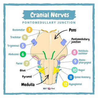
The facial nerve (VII) is a mixed one, considering its motor root in association with the sensitive root given by the intermediate (Wrisberg) nerve1. The taste of the anterior two-thirds of the tongue on each side are its responsibility. From the tongue, this afferent, pre-ganglionic route follows through the lingual nerve (association of nerves V and VII), and afterward through the tympanic cord nerve (facial branch), to make synapses on the geniculate ganglion. Through the intermediate nerve, the postganglionic fibers (afferent visceral special - gustative route) synapse in the solitary tract nucleus of the medulla oblongata, associated with the general afferent visceral fibers, providing sensitive innervation to the mucosa of the nasal cavities and soft palate.


The parasympathetic efferent fibers of the facial nerve, originating from the upper salivary nucleus located on each side of the upper portion of the medulla oblongata, run through the intermediate nerve and afterward through the tympanic cord nerve to make synapses in the submandibular ganglion. Thence, through postganglionic fibers, they stimulate salivary secretion of the submandibular and sublingual glands.


The motor portion of the facial nerve has its nucleus on the ventral portion of the pons. Its fibers stimulate the skin-inserted muscles in the face, neck, and scalp, as well as the posterior belly of digastric and stylohyoid muscles.


Symptoms of a NET
 experience the following tumor-related symptoms or signs:

My

Fatigue

Loss of appetite

Unexplained weight loss

Symptoms related to the tumor size and/or location:

Persistent pain in a specific area

Thickening or a lump in any part of the body

Nausea or vomiting

A cough or hoarseness that does not go away

Changes in bowel or bladder habits

Jaundice, which is the yellowing of the skin and whites of the eyes

Unusual bleeding or discharge

Symptoms related to the release of hormones:

Diarrhea

Facial flushing, usually without sweating

Hyperglycemia, which is a high level of glucose in the blood. Glucose is a sugar that is converted into energy by the body. Hyperglycemia causes frequent urination, increased thirst, and increased hunger.

Hypoglycemia, which is a low level of glucose in the blood. It causes fatigue, nervousness and shakiness, dizziness or light-headedness, sweating, seizures, and fainting.

Ulcer disease

Skin rash

Confusion

Anxiety

 The symptoms caused by a somatostatinoma may include the following:

pain in the abdomen (most common symptom)
diabetes
unexplained weight loss
gallstones
steatorrhea, or fatty stools
bowel blockage
diarrhea
 Peripheral effects of serotonin are local vasoconstriction where it is released and also vasodilatation and increased capillary permeability; constriction of veins and induction of venous thrombosis and promotion of platelet aggregation.

Serotonin has a positive chronotropic effect on the heart through 5HT4 receptors and can cause cardiac rhythm disorders.

• Excessive serotonin gives rise to debilitating diarrhea affecting the quality of life of patients with carcinoid syndrome.

• Prolonged exposure to high level of serotonin can cause fibrosis of heart valves, more commonly on the right side,

causing valvular heart disease. Excessive serotonin is also linked to fibrosis of uterus, skin (scleroderma),

pulmonary and retroperitoneal fibrosis in the long run,causing multiple complications.

Low levels of serotonin are often associated with many behavioral and emotional disorders.
10 months of diarrhea wore me out.
Maryfilomena E Rita Viñcenza I am honoured by your comment

"Payel Bhattacharya  you are what I call a beautiful soul . You see, I knew !!!! I see such light coming from you . There is a special reason why God saved you and uses you for his glory . I am so honored to have Met you . May God bless you . You have lightened up my mind and spirit ; you see , it is 20 years from 9/11, my love of my life Til death did we part , Mark , died from being a first responder a NYPD Sgt , who got kidney cancer from working months there . This puts my gift of life into perspective once again . I like you always chose to find the flowers the birds the breeze the music in life in the lil moments of the day . I have lost so much of my light since Mark died nearly 8 years ago . This is how I have written poems in the past and more importantly how I have spoken to encourage and enlighten others in their dark moments . Thank you . I’ll pray blessings over you . 💝"

I am happy to have a great friend too. I hardly got any good words or support from Indians
Friendship is one of life's greatest treasures.
Souls connected in space and time beyond any measure 

Even if we don't see each other for years

Emotions yet untold;

As time goes on, and bonds grow strong,

They will all unfold.

You are a great friend and I am happy. Pray I get over it


Next story would be of a metastatic up-to-date yet ignorance drips from it. 

After completing their medical degrees do they plunge into the world of unique diseases, keep themselves updated, keep track of all those maladies?


 Knowledge is power, ignorance about your disease will lead you nowhere. I have come across a few such patients with inadequate and volatile knowledge and they haven't survived after the second surgery. Ah! Don't look at me in dismay! 

If you have watched the '3 idiots' movie one of the Hindi movies I watched, I watch Tom Cruise, Robert Downey Jr, witty and action in English where things are on the edge but I liked this one. You can never learn from a book by memorizing it. Real knowledge is understanding the phenomenon. I grew up reading Carl Sagan and I admire science and I always have a question in my mind. Indians are always showing their true colours But I have never seen any other country so I don't know except whatsapp or zoom chat with them medical profession has many self-centred egoistic people that think they know everything and all patients should just shut up and be grateful and any questioning of their ability or judgement is rare... Who am I a mosquito but it's the goodness that makes the world spin. And we feel the bright sun will bring good tidings. There is positivity similarly there is negativity and both balance the world.


"Please block me..I will be very grateful. That gives u the option of unblocking me shud u ever need urgent help" said that abusive doctor.


We applaud them as they work for a patient to save a life

"I am a doctor - it's a profession that may be considered a special mission, a devotion. It calls for involvement, respect and willingness to help all other people."

But now doctors need patient's help to keep their observation, Reason, Human Understanding, knowledge because instead of serving patient as a physician keeping their dignity and pride in a lethal cancer the Unintelligible clamour created with ruthlessness.
services which will be purchased from them by health authorities and fund holding GPs through negotiated contracts. The hospital becomes a shop and ignorant doctors selling their services.
Few diseases of the present have little in common with the diseases of the past because of their rarity but like an owl, you have to pore over abstruse medical documents and make it your pastime with a great penchant for general science with an interest in medicine.

When you are bombarded with heavy-duty medical words of a mysterious sickness as your diagnosis by medical men who claim to possess full knowledge of those things don't become trifle too dreamy and treat their words as Gospels.

I think of the overwhelming despair of the medical practitioner roaring,

“You come and sit in my chair; let me go sit in your chair.”

Ego

Their impossibly bumptious opinionated ego deflates showing all the indications of superiority. He has earned his degree through remarkably well medical training going through the rigors of medical school, how can an inferior creature ask him questions?

This peculiar outbreak of blind rage and reprisal I faced in the unfunny days of my first brain tumor.

I spoke with authority because it was my body he would work upon and if he doesn't do a very careful job with his scalpel with full knowledge of what he was about to do then my fragile life bird would have flown away. I had the full right to know what was to be done to me.

After completing their medical degrees do they plunge into the world of unique diseases, keep themselves updated, keep track of all those maladies?

As I posted my first experience with a doctor 
We are the capable men we have a lot of ego

Who are you? A mosquito? You can't be bigger than my ego


Life taught

Heart locked

Long journey walked

Mind unshocked

WhatsApp/ Facebook/ calls blocked

Nothing to loose

The War lost my juice

Nothing to lose

Here was the untold talk


I have a lot of questions in my mind apart from abibiliophobia

A face reader said before liver transplant "you have so many questions in mind and such efforts you make to find them when Death visits you, you are going to ask who are you ?

Where did feelings go? when they disappeared? Did they leave a chemical trace somewhere our minds so that if we could look inside ourselves we would see via the patterns of neurons some of the important things that had happened to us in our lifetimes? 


So live in the moment
Not being influenced by any comment;
Enjoying the perfection of the magical moment
As stars comment
Burn out the galaxy bright
Hurtling towards the goal like a meteorite
Blazing your own trail

Nobody is immortal,
 Everyone has his own expiration date.
 The closer you get to death,
 The more intensely you live. 
Only when you are coping with a somewhat death sentence.

 You shall try to live a self-indulgent life before realizing that this does not fulfill the meaning of existence. 
That shouldn't happen in life.

A Warrior fights the battle of life ---
All its dreadful possibilities without a whit of fear 
Without thinking that it’s unfair. 
Before we are inevitably visited by death 
 Thrust to total chaos A warrior’s mind gracefully dances to the rhythm of the music of creation and life.
 Thus, A Warrior Dies Dancing, That’s Who I Am… 

The upshot is life belongs to us but we belong to death
 Meanwhile dance away the time you get with all your heart ❤️