

Ministry of Health and family Welfare, Government of India formulated a National Policy for Treatment of Rare Diseases (NPTRD) in July, 2017.
a) location - a disease which is uncommon in one country may be quite common in other parts of the world;
b) levels of rarity - some diseases may be much more rare than other diseases which are also uncommon; and
c) study-ability - whether the prevalence of a disease lends itself to clinical trials and studies.
However, it has been contested that disease prevalence alone may also not be an accurate basis for defining rare diseases, as it does not take into account changes in population over time. Hence, some have suggested that a more reliable approach to arriving at a definition could be based on the factors of –
a) location - a disease which is uncommon in one country may be quite common in other parts of the world;
b) levels of rarity - some diseases may be much more rare than other diseases which are also uncommon; and
c) study-ability - whether the prevalence of a disease lends itself to clinical trials and studies. This underscores the need for further research to better understand the extent of the existing diversity of definitions for rare diseases and to examine the scope of arriving at a definition, which is best suited to the conditions in India. It shall be done on a priority basis as soon as sufficient data is available. Steps have already been taken for creation of a hospital based National Registry for rare diseases in India by ICMR.
Challenges in research and development
A fundamental challenge in research and development for the majority of rare diseases is that there is relatively little known about the pathophysiology or the natural history of these diseases. Rare diseases are difficult to research upon as the patient pool is very small and it often results in inadequate clinical experience. Therefore, the clinical explanation of rare diseases may be skewed or partial. The challenge becomes even greater as rare diseases are chronic in nature, where long term follow-up is particularly important. As a result, rare diseases lack published data on long-term treatment outcomes and are often incompletely characterised.
This makes it necessary to explore international and regional collaborations for research, collaborations with the physicians who work on any rare disease and with patient groups and families dealing with the consequences of these disorders. This will help gain a better understanding of the pathophysiology of these diseases, and the therapeutic effects that would have a meaningful impact on the lives of patients. There is also a need to review and where possible modify, clinical trial norms keeping in mind the particular challenges in rare diseases, without compromising on the safety and quality of
he drugs or diagnostic tools.
Mr. Narendranath Vikkath, biotechnologist and lecturer at Amrita institute of medical sciences said,
"In India we don’t have data i guess. May be you can get individual case reports"
Other names of VHL gene
- elongin binding protein
- pVHL
- VHL1
- VHL_HUMAN
- von Hippel-Lindau tumor suppressor, E3 ubiquitin protein ligase
Mutations in the VHL gene have been identified in a type of tumor called a hemangioblastoma. These tumors are made of newly formed blood vessels and tend to develop in the brain and spinal cord.
- Anesthesia for Emergency Craniotomy in a Patient with von Hippel Lindau Disease With Pheochromocytoma To JNA Readership:
von Hippel Lindau (VHL) disease is a rare autosomal dominant disorder characterized by the presence of hemangioblastomas in the brain, spinal cord, and retina with associated involvement of the kidneys, liver, pancreas, and adrenals.The presence of pheochromocytoma in a patient with VHL poses a considerable challenge to the anesthesiologist. This is a report of a patient with VHL and underlying pheochromocytoma who underwent emergency craniotomy for excision of a cerebellar hemangioblastoma.
A 36-year-old male patient was admitted to the emergency room with signs and symptoms suggestive of intracranial hypertension. He had a history of accelerated hypertension (230/120 mm Hg) 3 years earlier and was on regular medication. At the time of admission, his pulse rate was 110 per minute, and blood pressure was 170/110 mm Hg.
Fundus examination revealed an angiomatous malformation in the left eye.
MRI of the brain showed right cerebellar hemangioblastoma with mass effect and obstructive hydrocephalus. CT of the abdomen revealed bilateral suprarenal masses with multiple renal cysts. Raised vanillylmandelic acid-end stage metabolite of chatecholamines (154 mg, normal <13 mg) confirmed the presence of coexisting pheochromocytoma, and a diagnosis of VHL was made. Decongestive therapy was started to stabilize the neurologic status. He was planned for simultaneous craniotomy and bilateral adrenalectomy after adequate preparation with beta blockers. However, on the third day, sudden neurologic deterioration necessitated emergency craniotomy.
Intraoperatively, propofol, esmolol infusion, and sevoflurane (2%3%) were used to control hypertension and tachycardia, and he was extubated uneventfully using labetalol 0.1 mg/kg. In the postoperative period, his BP was controlled with nitroglycerin and esmolol infusion. Ten days later, he underwent bilateral adrenalectomy under combined general and epidural anesthesia. His postoperative period was uneventful, and he was discharged without any neurologic deficit.
Neurologic symptoms in a patient with VHL include posterior fossa signs,raised intracranial pressure (ICP), and spinal cord syndromes. Associated pheochromocytoma renders them prone to hemodynamic fluctuations, which can be hazardous because it can lead to raised ICP and intratumoral bleed. Therefore,adequate preparation with a and b blockers is essential for maintaining hemodynamic stability. In addition, availability of better anesthetic agents and invasive monitoring during the intraoperative and postoperative period has remarkably reduced pheochromocytoma-related mortality from approximately 70% to less than 3%.2
Because this patient was taken up for craniotomy as an emergency, medical optimization was not possible, and hemodynamic fluctuations, especially during intubation and extubation, were anticipated. Vasodilators such as nitroglycerin,3 sodium nitroprusside, and in-halational agents have the potential to increase cerebral blood flow and ICP in patients with intracranial hypertension; therefore, esmolol and propofol were preferred until dural opening. Later, sevoflurane was used because it provides tighter control of blood pressure in pheochromocytoma.Towards the end of surgery, sevoflurane was discontinued to facilitate rapid awakening and early extubation, and infusions of nitroglycerin and esmolol were started to maintain hemodynamics. This was continued in the postoperative period along with invasive monitoring to enable titration tion of blood pressure to acceptable levels.
Thus, optimization of the preoperative status along with tight control
of hemodynamics perioperatively with suitable drugs, and invasive monitoring can lead to a good outcome in such high-risk patients.
Sanjay Goel, MD
Neerja Johar, MD
Mary Abraham, MD
Department of Anesthesiology
Fortis Hospital
Noida, India
Central nervous system hemangioblastomas are cardinal
feature of VHL syndrome and occur in 60-80% of VHL
patients, with cerebellum being the most common
site . VHL syndrome associated hemangioblastomas
frequently expresses SSTR . Ambrosini et al. have
previously demonstrated in vivo SSTR expression in VHL
associated hemangioblastoma with 68Ga-DOTANOC PET-
CT. In the present case 68Ga-DOTANOC PET-CT detected
previously unknown cerebellar hemangioblastoma, which
was confirmed on contrast enhanced MRI. Retinal angiomas
(hemangioblastoma) are the most common presenting
feature of VHL disease as was in the present case, though
not recognized at that point of time. 68Ga-DOTANOC PET-
CT detected the retinal lesions and were subsequently
confirmed with MRI. To our knowledge, there is no previous
published report of imaging retinal hemangioblastoma with
68Ga-DOTANOC PET-CT.
~ Von Hippel-Lindau Syndrome: Demonstration of Entire Disease Spectrum with 68Ga-DOTANOC PET-CT Punit Sharma, MD, Varun Singh Dhull, MD, Chandrasekhar Bal, MD, Arun Malhotra, PhD, Rakesh Kumar, MD, PhD All authors: Department of Nuclear Medicine, All India Institute of Medical Sciences, New Delhi 110029, India
Von Hippel-Lindau disease – A case report
G. Venkatesh
Chennai Medical college Hospital and Research
Centre, Trichy, Tamil Nadu, India
Introduction: Von Hippel-Lindau (VHL) disease is a rare multis-system disorder with autosomal dominant inheritance presenting with visceral cysts and benign tumors involving multiple organs.
The spectrum of presentation of disease includes Retinal and Central Nervous System Hemangioblastoma, Pancreatic cysts and tumors, renal cysts and tumors etc. The diagnosis of disease remains challenging because of its varied age presentation and protean (variable) nature of clinical manifestation. Imaging plays a pivotal role in the diagnosis and follow ups of VHL patients. Early diagnosis and follow up of VHL disease is important due to the risk of malignant transformation of renal cyst.
Case report: A 21 year old female presented to neurosurgery as known case of brain tumor (medulloblastoma with spinal metastasis). The patient was referred to radiology for routine preoperative imaging evaluation. Ultrasound imaging showed pancreatic cysts at head and tail region. Spinal magnetic resonance imaging (MRI)was done which showed multiple hemangioblastoma. Though the clinical presentation pertained to a single system initially, the imaging of abdomen and brain helped to clinch the diagnosis as VHL disease.
Conclusion: A review of the genetic basis of VHL disease in patients will be discussed. Emphasis should be made on importance of annual clinical screening of the patient and genetic screening of at-risk family members.
About 60%–80% of subjects with von Hippel Lindau (VHL) syndrome develop central nervous system (CNS) hemangioblastomas, which are vascular tumors composed of a thick network of blood vessels and pericytes (VHL registry, UK). The VHL protein forms heteromeric complexes with Elongin B, Elongin C and Cullin proteins that bind to HIF-1α on its hydroxylated proline residues to direct a regulated proteolysis of HIF-1α. At lower oxygen concentrations, the regulated hydroxylation of prolines rescues HIF-1α to induce a set of hypoxia-inducible genes, such as vascular endothelial growth factor (VEGF), Glucose transporter-1 (GLUT1) and Erythropoietin (EPO) , which cause endothelial cell proliferation and neovascularisation. In the VHL syndrome, the lack of a functional VHL protein attributed to the truncating or disruptive heritable mutations of the VHL gene leads to the uninhibited HIF-1α directed induction of hypoxia-inducible genes which, in turn, is thought to promote angiogenesis and, ultimately, hemangioblastoma. However, we and others have shown from germline mutational analysis that many disruptive mutations of the VHL gene would not necessarily lead to HB .
Several causative factors for the hypervascularization of the extracellular matrix (ECM) have been implicated.
Fibronectin (FN) is expressed abundantly in the vascular matrix of embryonic tissues and remains in the basement membrane matrix of blood vessels throughout life.
(Fibronectin (FN) is a high-molecular-weight glycoprotein component of the extracellular matrix involved in cell adhesion, migration, metastasis, proliferation and differentiation, as well as embryogenesis, wound healing, and blood coagulation. Considerable recent research has established that tumor expression of FN is closely associated with tumor formation and development as well as disease prognosis. However, the mechanisms underlying this relationship have remained unclear. The aim of this study was to investigate FN protein expression in esophageal squamous cell carcinoma (ESCC) and determine its potential prognostic relevance, while also elucidating the source and function of FN.
~ Expression of fibronectin in esophageal squamous cell carcinoma and its role in migration
Jiefei Xiao, Weilin Yang, Bo Xu, Haoshuai Zhu, Jianyong Zou, Chunhua Su, Jian Rong, Tao Wang & Zhenguang Chen
BMC Cancer volume 18, Article number: 976 (2018) Cite this article)
The cross-linking of soluble FN after an intracellular signaling-regulated priming and disulfide linkages results in a detergent-insoluble matrix form of FN. The matrix form of FN is critical for tissue scaffolding in endothelial cell-initiated angiogenesis. Moreover, FN deposition and matrix formation are critical for the downregulation of the integrin assembly in renal cell lines. A significant role played by FN protein deposition and cross-linking in the ECM has been associated with VHL protein.However, the role of FN in HB has not been studied previously.
The current study was performed to understand the role of FN in the pathogenesis of VHL-associated HB. A comparative expression pattern of FN and VHL in HB was done using in vitro biological functional tube formation assay using specific FN substrates and inhibitors on endothelial cells obtained from human umbilical cord.
Several studies have shown an association between VHL and FN during embryogenesis as well as tumorigenesis.
Furthermore, we demonstrated that the VHL syndromic hemangioblastomas have very low FN expression compared to the control tissues. We have also shown that FN is expressed moderately in the molecular layer of normal cerebellar tissues in IHC studies. A recent study has shown that the VHL gene knock down in endothelial precursor cells called ‘hemangioblasts’ causes the development of hemangioblastoma and retinal angioma but not RCC in mice. Therefore, the loss of the VHL protein in endothelial cells causes the abnormal angiogenesis and eventual HB tumorogenesis via the defective matrix FN deposition. VHL is a known tumor suppressor protein and the tumors associated with the VHL syndrome are hypervascular in nature despite being benign . Studies have demonstrated the direct role of VHL in downregulating HIF-1α and in controlling the hypoxia dependent survival mechanism . The key cell types initiating the neovascularisation process are endothelial cells. In order to understand the HIF-1α-independent mechanism of VHL in controlling vasculogenesis, we used the endothelial tube formation assay as our experimental model and found that the VHL protein expression is closely connected with the tube formation dynamics of the endothelial cells. Additionally, we were able to demonstrate an incremental pattern of FN deposition by the endothelial cells during the tube formation process. Experiments in which FN was added externally in both its plasma and matrix forms showed that soluble plasma FN upregulated the tube formation process, while the matrix form of FN inhibited tube formation.VHL is known to have roles in extracellular FN matrix assembly. VHL knockout cell lines have defective matrix FN assembly, which has been demonstrated in clear cell RCC (ccRCC) associated with VHL . A study has shown that a mutant VHL protein, despite being able to suppress HIF-1α but lacking the ability to deposit matrix FN, did not inhibit tumor growth . This implicates the crucial role of the defective matrix FN assembly function of VHLrelated angio- and tumorigenesis.
FN is seen throughout the molecular layer of cerebellar tissue and is abundantly seen in vascular development. During the neovascularisation process, FN is expressed in the mesenchyme and stays at the basement membrane surrounding the blood vessels. It has been shown that FN null endothelial cells make blood vessels dilate due to the lack of contact with the mesenchyme . Several studies have shown the role of FN matrix assembly in endothelial cells during blood vessel formation, definitely pointing towards the crucial role played by endothelial cells in converting soluble FN into matrix form . It also emphasizes the fact that defective matrix FN in the basement membrane of endothelial cells leads to defective blood vessel formation and unchecked angiogenesis. In this study, we demonstrate that VHL-associated hemangioblastomas have low expression of FN in the tissue; a phenomenon that is related to the absence of VHL in the endothelial cells of the tumor vascular structure. By immunohistochemistry studies, we have identified that FN is expressed moderately in the molecular layer of the normal cerebellum but absent in the VHL syndromic hemangioblastomas. This suggests that the VHL-mediated deposition of FN in the basement membrane of blood vessels in the cerebellum plays a crucial role in hemangioblastoma development. The ECM in blood vessels plays a critical role in vessel integrity. Studies have demonstrated the leaky nature of vascular structures in VHL null endothelial cells, thus emphasizing the FN-dependent HIF-1α independent functionality of VHL .
Furthermore, we have shown that a cyclic RGD peptide inhibited the endothelial tube formation process. During the matrix deposition, FN changes its structure and exposes RGD groups hidden in the 3D structure of the protein, which in turn, attaches with the cell surface integrin receptors to execute its functions. Therefore, the RGD peptide moiety of FN, which becomes actively exposed during the 3D structural changes occurring during its matrix deposition perhaps act as an important angiogenesis switch that is defective in the VHL disease due to the lack of VHL protein function . In the endothelial cells, a timely expression of VHL is needed for that FN is expressed moderately in the molecular layer of the normal cerebellum but absent in the VHL syndromic hemangioblastomas. This suggests that the VHL-mediated deposition of FN in the basement membrane of blood vessels in the cerebellum plays a crucial role in hemangioblastoma development. The ECM in blood vessels plays a critical role in vessel integrity. Studies have demonstrated the leaky nature of vascular structures in VHL null endothelial cells, thus emphasizing the FN-dependent HIF-1α independent functionality of VHL .
Furthermore, we have shown that a cyclic RGD peptide inhibited the endothelial tube formation process. During the matrix deposition, FN changes its structure and exposes RGD groups hidden in the 3D structure of the protein, which in turn, attaches with the cell surface integrin receptors to execute its functions [5]. Therefore, the RGD peptide moiety of FN, which becomes actively exposed during the 3D structural changes occurring during its matrix deposition perhaps act as an important angiogenesis switch that is defective in the VHL disease due to the lack of VHL protein function. In the endothelial cells, a timely expression of VHL is needed for well-regulated vasculogenesis to occur. In our study, we have shown that the functional RGD moiety has the ability to inhibit the endothelial tube formation process. Previous studies have demonstrated the antiangiogenic and antitumorigenicpotential of matrix FN . We believe this is an important finding as cilengitide – a cyclic RGD peptide already tested as an anti-angiogenic drug in clinical trials for high-grade glioma – may be utilized for this target for the treatment of prevention of HB formation in VHL patients.
Author contributions: All the authors have accepted responsibility for the entire content of this submitted manuscript and approved its submission.
Research funding: Indian Council of Medical Research, Funder Id: 10.13039/501100001411, Grant Number: 54/18/2011-HUM/BMS.
Employment or leadership: None declared.
Honorarium: None declared.
Competing interests: The funding organization(s) played no role in the study design; in the collection, analysis, and interpretation of data; in the writing of the report; or in the decision to submit the report for publication.
~ Narendranath Vikkath*, Prasanth Ariyannur, Krishnakumar N. Menon, Bindhu MR and Ashok Pillai*
Exploring the role of defective fibronectin matrix assembly in the VHL-associated CNS hemangioblastomas
This ICMR has funded research on von Hippel-Lindau which is a genetic cancer.
The unusual clinical features of this disorder predicted a role for the von Hippel–Lindau gene (VHL) in the oxygen-sensing pathway. Indeed, recent studies of this gene have helped to decipher how cells sense changes in oxygen availability and have revealed a previously unappreciated role of prolyl hydroxylation in intracellular signaling. These studies, in turn, are laying the foundation for the treatment of a diverse set of disorders, including cancer, myocardial infarction, and stroke.
Different VHL types and subtypes can be distinguished based on predominant pathology in reference to pheochromocytomas, as follows:
VHL type 1 - Renal carcinoma and hemangioblastoma.
VHL type 2A - Hemangioblastoma and pheochromocytoma.
VHL type 2B - Renal carcinoma and pheochromocytoma.
VHL type 2C - Pheochromocytoma only.
VHL is a tumor-suppressor gene located on the short arm of chromosome number 3. It is widely expressed in normal fetal and adult tissues, yet mutations in VHL leads to development of various human neoplasms including renal cell carcinoma.It has been proven that familial as well as sporadic renal cell carcinomas are associated with VHL gene mutations.
The VHL gene produces proteins of 213 and 160 amino acids, respectively.These proteins display similar biochemical properties and both forms are referred to as pVHL, VH protein or VHL gene product. This VHL protein plays important role in cell cycle regulation and acts as tumor suppressor product of VHL gene. Mutations in VHL gene leads to structural alteration in VHL protein predisposing to development of renal cell carcinomas. VHL gene mutations in renal cell carcinoma have been studied widely at cytogenetic and molecular level in both familial as well as sporadic renal cell carcinomas but there are very few studies that analyzed association of VHL and renal cell carcinomas at protein level. To examine VHL expression at the protein level, we have performed an immunostaining of renal cell carcinoma specimen tissues using monoclonal antibodies to pVHL.
In present study we evaluated 32 cases of renal cell carcinoma for pVHL expression and We found that pVHL expression was positive in 84.40% (n=27) of cases of renal cell carcinoma and rest of 15.60% (n=5) cases were found to be negative for it. Our findings are in agreement with a recent study by Schraml et al on analysis of pVHL expression in renal cell carcinoma by immunohistochemistry using poly and monoclonal antibodies against VHL protein. They found that immunohistochemically, pVHL was expressed in 70% of cases of RCCs under their study and rest 30% was found to be negative for VHL protein.
Lin et al immunohistochemically evaluated renal and nonrenal tumors for usefulness of pVHL by using rabbit polyclonal antibodies against VHL protein. They concluded strong positive immunoreactivity for pVHL and nearly 100% primary renal cell carcinomas and 95% of metastatic RCCs showed positive immunoreactivity for VHL protein. Our results are supported by study of Corless et al who had immunohistochemically detected VHL protein in 80% cases of RCCs and rest were negative for it . They also gave a very important statement regarding negative staining of pVHL in renal tumors. They stated that mutated VHL gene is predicted to produce altered forms of VHL protein that is truncated or elongated with respect to Elongin BoC binding region. These mutant forms of VHL protein should be remain detectable by immunostaining provided that the appropriate epitopes remain intact . Schraml et al , also reported that allelic deletion of the VHL gene was not associated with pVHL expression. Schumeli et al stated that loss of function mutations in the Von Hippel-Lindau (pVHL) tumor suppressor protein are tumorogenic and by using biophysical methods confirmed that mutant pVHL proteins have lower stability than the wild type, distorted core domain and as a result reduce the ability of the protein to bind its target HIF-1α.
Importantly, all recent studies of human tissues have exclusively described cytoplasmic pVHL expression. Only some immune fluorescence analyses have shown that pVHL can be expressed, to a lesser extent, in the nucleus or in association with cell membranes. Only few recent studies have examined the combined cytoplasmic and nuclear localization of VHL protein in RCCs and its clinical relevance. In present study, among all pVHL positive cases, combined cytoplasmic and nuclear expression of pVHL was most common (59.0%) followed by only cytoplasmic expression (37%). Nuclear expression alone was rare and was noted in only one case. Our this finding is in agreement to the study of Schraml et who found combined cytoplasmic and nuclear expression of pVHL was commonest and nuclear alone was rarest pattern in RCCs.
We analyzed pVHL expression in different histological subtypes of RCCs. Positive pVHL expression was found in 91.30 % of clear cell RCCs, 75 % of papillary RCCs .Only one case of chromophobe RCC was found in our study, that came to be negative for VHL protein expression. Schraml et detected VHL protein expression in 69 % clear cell carcinoma, 68 % papillary carcinoma and 76% of chromophobe carcinomas while Lin et found immunohistochemica positivity for VHL protein in 99% clear cell RCC, 100 % papillary and 100 % chromophobe RCCs. This discrepancy in findings may be attributed to different methods of immunostaining and different kind of antibodies used for detection of pVHL expression.
Among clear cell RCCs, 47.80% showed combined cytoplasmic and nuclear pVHL expression, 39.10% showed only cytoplasmic expression and 4.30% showed only nuclear expression. Out of all pVHL positive papillary RCCs, 62.50% showed combined cytoplasmic and nuclear expression, 12.50 % showed cytoplasmic expression and no isolated nuclear expression was seen. Hence, we concluded that combined cytoplasmic and nuclear expression of pVHL is most common pattern of expression in RCCs. Schraml et al found 44% clear cell RCCs and 44 % of papillary RCCs were positive for combined cytoplasmic and nuclear expression. We observed that exclusive nuclear expression of pVHL was the least common pattern but in contrast to our finding they observed exclusive cytoplasmic expression as least common pattern.
~ VHL protein expression in renal cell carcinoma
Yadav R 1, Sagar M 2, Kumari M 3, Singhai A 4, Babu S 5, Kumar A 6
1Dr Rita Yadav, Senior Resident, 2Dr Mala Sagar, Associate Professor, 3Dr Malti Kumari, Associate Professor, 4Dr Atin Singhai, Associate professor, 5Dr Suresh Babu, Professor, 6Dr Ashutosh Kumar, Professor & Head, All are affiliated with Department of Pathology, King George Medical University, Lucknow, India.
Glioblastomas (GBM) are the commonest and most lethal intracranial tumours. The GBMs exhibit a relentless malignant progression characterized by invasiveness and resistance to therapy.In GBM, hypoxia is readily recognizable, resulting in the necrotic areas in the core of the tumour and surrounded by evidence of hypoxic response and neovascularisation. It is known that hypoxia contributes to an aggressive phenotype in tumours, including features of resistance to apoptosis, drug resistance, increased invasiveness and tumour heterogeneity and aggressive growth patterns. Survival and proliferation of GBM cells under such adverse condition requires an adaptation to the environment, wherein, hypoxia‐inducible factor‐1 is a key adaptive molecule for regulating the cellular response. On activation and stabilisation during hypoxia, HIF1α upregulates target genes involved in the process of angiogenesis, tumour invasion and metastasis, energy metabolism, and adaptive survival in GBM,and tumours at‐large. Overexpression of HIF1α in surgically resected tumour specimens has been shown to have positive correlation with tumour aggressiveness and poor prognosis in different tumours including GBM. HIF1α is known to be regulated by growth factors,cytokines and mitogens. pathway is a known positive regulator of HIF1α and this pathway may function via mTOR or independent of it The EGFR/Akt pathway is known to be frequently activated in GBM.
Taken together, the results indicate that at least in glial tumours with upregulated FAT1, there is a major role of this atypical cadherin in the upregulation of HIF1α during severe hypoxia. This further affects metabolic adaptation and augments the invasive properties of the tumour cells. On the basis of these findings we conclude that FAT1 is a novel regulator of HIF1α under severe hypoxia via both EGFR/Akt/mTOR axis (synthetic) and VHL (proteasomal degradation) in GBM. This is the first report on the important role of FAT1 in modulating HIF1α expression and activity representing a novel pathway, essential for glioma progression and aggressiveness. This study raises the possibility of FAT1 acting as a regulator of the master regulator HIF1α and forming a link between hypoxia and cancer in glioma. This along with the positive association observed between FAT1 and HIF1α in GBM tumour tissues which initially suggested the regulatory role of FAT1 points to a clinical relevance for this cadherin in those GBMs where it is upregulated. While we have studied this pathway only in glioma, it is possible that this FAT1‐HIF1α axis is relevant in other tumour types as well. Till now, targeting HIF1α or other hypoxia markers/pathways as therapeutic strategies to treat cancers has had a limited success.49 The FAT Atypical Cadherin could possibly be a novel target for therapeutic intervention in gliomas with high FAT1 expression and hypoxic microenvironment.
*FAT1 an essential role for cellular polarization, directed cell migration and modulating cell-cell contact.
*HIF1α-is known to induce transcription of more than 60 genes, including VEGF and erythropoietin that are involved in biological processes such as angiogenesis and erythropoiesis, which assist in promoting and increasing oxygen delivery to hypoxic regions.
*EGFR/Akt- Epidermal growth factor (EGF) binding to its receptor (EGFR) activates several signaling intermediates, including Akt, leading to control of cell survival and metabolism.
*mTOR- mechanistic target of rapamycin{Sirolimus, also known as rapamycin}(mTOR)
*Cadherins are transmembrane proteins that mediate cell–cell adhesion in animals. By regulating contact formation and stability, cadherins play a crucial role in tissue morphogenesis and homeostasis
After several misdiagnosis and diagnostic dilemmas in 2008 I was diagnosed with a very rare disease hardly known in India, called VHL or von Hippel Lindau during my landmark liver transplant. I can still recall the faces of the radiologists checking my tumour studded liver with their probes and looking at my file with a weird name “VHL” with vacant faces and regarding me quizzically. Yes, the liver transplant was a life and death affair and we had no money to get the liver transplant done which was a whopping> 30 lakhs!
But now all the above data and more I have come across as a patient says that not only doctors who have treated me but all radiologists are aware of the disease.
At that time liver and lung lesions weren't found in the USA but now the scene has changed.
Lots of mew data has evolved and the VHL gene is behind a wide range of cancers. Treating VHL Is one step closer to treating cancer.
I was diagnosed with a rare disease von Hippel-Lindau or VHL which is a genetic defect that causes capillary growth to go out of control. While the tiniest blood vessels or capillaries usually branch out gracefully like trees, in VHL patients a little knot of extra capillaries forms a growth or tumour and in certain cases, it turns cancerous. It is a genetic form of cancer VHL patients battle a series of tumours throughout their life. VHL may occur in up to 10 organs of the body like liver, kidney, brain, spinal cord or retina, inner ear, pancreas, pheochromocytoma, paraganglioma, neuroendocrine tumours can also happen.
Treatment for VHL varies according to the location and size of the tumour. In general, the objective of treatment is to treat the tumours before they grow to a size large enough to cause permanent problems by putting pressure on the brain or spinal cord. Treatment of most cases of VHL usually involves surgery to remove the tumours before they become harmful. Certain tumours can be treated with focused high-dose irradiation. Individuals with VHL need careful monitoring by a physician familiar with the disorder/ treated or familiar with this patient's case.
I have faced countless surgeries and there will be more on the way.
Trigeminal neuralgia is an extremely debilitating condition unlike many other medi-
cal ailments. The paroxysmal pain is so unbearable that it completely breaks down
the mental strength of a person, sometimes to the point of driving him/her to even attempting suicide. My mentor, Prof. Parmod Kumar Bithal, could realise my growing interest in this particular group of patients. As a senior faculty and head of the department, Dr. Bithal gave me a lot of liberty to execute treatment plans with his constant encouragements. A lot of discussions with him further motivated me to participate regularly in treatment strategies for patients with trigeminal neuralgia, who were referred to us by colleagues from the specialities of neurology and neurosurgery. The intention to do something more for these patients fuelled my desire to read more and get updated with relevant literature.
I convey my heartfelt gratitude to all the authors and coauthors, who contributed immensely with lots of information on their respective topics. I am thankful to the director of AIIMS, Prof. Randeep Guleria, for permitting me to edit this book. I take this opportunity to thank my beloved teacher, Prof. Hari Hara Dash, for being the constant source of encouragement during my academic journey.
~
Prof. Girija Prasad Rath of the All India Institute of Medical Sciences (AIIMS), New Delhi.
Epidemiology The overall incidence of TGN is about 40–50 cases per one million and estimated prevalence is approximately 100–200 per million population. Incidence of TGN varies signiicantly with age, with less than 5 per million in people younger than 18 years, and some studies estimating as high as 800 per million in older age groups . Patients in the age range of 35–65 years are most commonly affected. For reasons that are not properly understood, TGN is nearly twice more common in females than males. Hereditary forms of TGN have been reported but are rare and constitute less than 4–5% of overall TGN . However, patients with bilateral TGN have a higher hereditary predisposition than those with unilateral presentation. Although there is no clear evidence, people with hypertension and migraine seem to be at an increased risk of developing TGN.
~ Handbook of Trigeminal Neuralgia
Not going into further details kindly note Prof. Randeep Guleria had treated my MDR Tuberculosis for 2 years and
Prof. Hari Hara Dash had treated my TGN for a brief time at Fortis Memorial Research Institute Gurugram
Hypoparathyroidism is an uncommon disorder of
calcium metabolism characterized by hypocalcemia,
hyperphosphatemia, and reduced level of intact parathyroid
hormone (iPTH).
An amide of PTH, is widely available for the use in osteoporosis; however, its use in hypoparathyroidism is limited.
Aims: The aim of this study is to evaluate the efficacy of PTH (1–34) in the treatment of patients with hypoparathyroidism.
Settings and Design: This was a prospective, open‑label interventional study in a tertiary care hospital of Indian Armed Forces.
Subjects and Methods: All patients with hypoparathyroidism presented to the endocrinology outpatient department were included and were exhibited injection PTH (1–34) 20 µg twice daily that was gradually reduced to 10 µg twice daily along with calcium, active Vitamin D (alfacalcidol), and hydrochlorothiazide. Oral calcium and alfacalcidol doses were also reduced to maintain serum calcium within normal range. The quality of life (QOL) score was calculated using RAND 36 QOL questionnaire at baseline and termination of the study.
Statistical Analysis Used: Paired t‑test was used to calculate pre‑ and post‑treatment variables.
Results: Eight patients (two males) were included in this study having mean age of 35.8 years. PTH (1–34) treatment led to the improvement in serum calcium (6.81–8.84 mg/dl), phosphorous (5.8–4.2 mg/dl), and 24 h urinary calcium excretion (416–203.6 mg). Parameters of QOL showed the improvement in overall QOL, physical performance, energy, and fatigue scores. No major adverse events were noted.
Conclusions: Treatment of hypoparathyroidism with PTH (1–34) leads to improvement in calcium profile, reduction in hypercalciuria, and improvement in QOL, whereas it is safe and well tolerated.
Keywords: Hypoparathyroidism, quality of life, teriparatide
~ Efficacy of Teriparatide in Patients with Hypoparathyroidism: A Prospective, Open‑label Study Vimal Upreti, Shrikant Somani, Narendra Kotwal Department of Endocrinology, Army Hospital (R and R), New Delhi, India
Indian journal of anaesthesia and Indian journal of endocrinology and metabolism contains few (10)such articles.
I had a subtotal thyroidectomy in 2006 for multi-nodular thyroid leading to goitre with retro-sternal extension. It was one of the longest surgeries lasting for 6 whole hours.
In a routine ultrasound (after the lymphadenopathy surgery as prescribed by Dr. Randeep Guleria) of the neck on May 14th 2014, it was found…
"Left lobe of thyroid is not visualised (postoperative).
Remnant of the right lobe measures 5.2x8.9 mm. Adjacent to it at least 4 hypoecohypoecoic nodules seen measuring 6.2 mm, 10.8 mm, 11.5mm, and 12 mm in size with increased vascular flow within as well as at the periphery on CDF1 images. These are suggestive of hemangioblastomas in a known case."

It is critically important for the thyroid surgeon to employ strategies for minimizing and preventing hypoparathyroidism (or Hypopara for short), including carrying out the most appropriate extent of thyroidectomy for a specific patient.
Hypocalcemia causes
neuromuscular excitability and cardiac electrical instability due to a reduced nerve and muscle cell depolarization threshold.
It's most common early symptoms are paresthesias, or numbness and tingling, of the perioral region and the fingertips. Muscle stiffness, cramps, and spasms are also common.
Neuropsychiatric symptoms include confusion, anger, depression, lightheadedness, and irritability. More sustained muscle contraction may lead to laryngospasm, and more severe neural excitability may lead to seizures.
Signs of hypocalcemia include observed or elicited tetany.
Classic bedside findings are a positive Chvostek sign (facial muscle twitching upon tapping the preauricular region over the facial nerve; present at baseline in up to 25% of people), or a positive Trousseau sign (flexion of the wrist, thumb, and metacarpophalangeal joints and hyperextension of the fingers, upon brachial artery occlusion by inflation of a blood pressure cuff above systolic blood pressure).
Cardiovascular signs observed with progressive hypocalcemia include prolongation of the QT interval that can result in torsades de pointes, a form of ventricular tachycardia that may degenerate into ventricular fibrillation.
Insufficient PTH leads to low calcium levels in the blood, or hypocalcaemia. It causes electrolyte imbalance and can be a life-threatening condition if untreated.
Why is calcium so important? Calcium is vital to life and affects every cell in the body. Most people know about teeth and nails in connection with calcium but its effects are on the whole body - nerves, muscles, and organs. It helps blood to clot and is important in energy production. Calcium is crucial to us which is why the body has special mechanisms like the parathyroid glands to keep calcium levels constant.
Treatment with vitamin D analogues and calcium supplements is not ideal and can lead to long term renal problems. Calcium levels fluctuate but home calcium testers are not available so monitoring this condition can be challenging. Until 2015, Hypopara was the only endocrine condition without its own replacement hormone. Injectable parathyroid hormone is now licensed for use in the treatment of hypopara in the USA but is not yet available elsewhere.
But as I see in Army hospital parathyroid injections are available not for civilians
I get treatment of my HypoPara with calcitriol and calcium by Endocrinologist Dr. Ambrish Mithal a year ago even after partial nephrectomy my liver and kidney were functional yet now we are uncertain due to the pandemic and being strapped for funds many tests haven't been done
What is an orphan drug?
"Orphan drugs" are medicinal products intended for diagnosis, prevention or treatment of life-threatening or very serious diseases or disorders that are rare.
An orphan drug is a pharmaceutical agent developed to treat medical conditions which, because they are so rare, would not be profitable to produce without government assistance. The conditions are referred to as orphan diseases.
Jan. 9, 2006 — The FDA has approved orphan drug status for mycophenolate mofetil (a bottle is very expensive in India and you can use it for 60 days then you have to throw it away)
Mycophenolate is approved by the FDA and other regulatory authorities worldwide for use in conjunction with cyclosporine( Neoral which I had to quit because of gingival hyperplasia) and corticosteroids which caused posterior subcapsular cataracts….
for the prophylaxis of organ rejection in adult patients receiving allogeneic renal, cardiac, or hepatic transplant. In some countries, it is also approved for use in pediatric kidney transplantation.
Liver transplantation is an important treatment option for selected patients with non-resectable multiple tumors.
When multiple tumors formed in the liver which compressed and displaced vital vessels and I had a liver transplant.

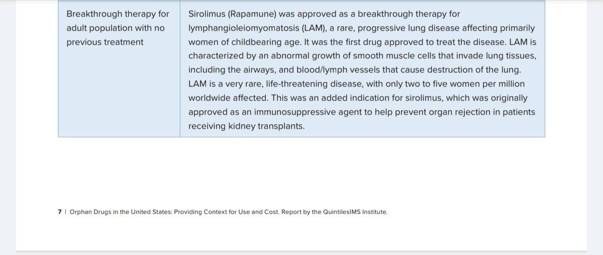
Sirolimus is a natural macrocyclic lactone(are products or chemical derivatives of soil microorganisms belonging to the genus Streptomyces)produced by the bacterium Streptomyces hygroscopicus, with immunosuppressant properties. In cells, sirolimus generates an immunosuppressive complex that binds to and inhibits the activation of the mammalian Target Of Rapamycin (mTOR), a key regulatory kinase( Kinase, an enzyme that adds phosphate groups (PO43−) to other molecules. A large number of kinases exist—the human genome contains at least 500 kinase-encoding genes.). This results in inhibition of T lymphocyte activation and proliferation that occurs in response to antigenic and cytokine stimulation and inhibition of antibody production.
Sirolimus can prevent angiogenesis by interfering with vascular endothelium growth factor (VEGF)‐mediated pathways in endothelial cells, thus limiting the growth of tumors,and also impacts established tumors, by inducing extensive microthrombi and so inhibiting tumor growth.

My orphan drugs supplied by https://www.panaceabiotec.com/about-panacea-biotec
No comments:
Post a Comment